

For generations, dental visits were associated with high anxiety, physical discomfort, and prolonged treatment timelines. The mere sound of a high-speed drill or the prospect of biting into a tray filled with cold, sticky impression putty was enough to make many individuals delay essential oral healthcare.
The dental landscape has undergone a profound shift. The integration of advanced digital systems, advanced imaging, bioprinting, and minimally invasive tools has fundamentally changed the nature of clinical treatment. Modern dentistry focuses heavily on precision, customization, and patient comfort. These clinical innovations streamline complex procedures, optimize treatment accuracy, and significantly alleviate the stress historically linked to the dental chair.
Digital Impressions and Intraoral Scanners
One of the most immediate changes patients notice in a contemporary dental office is the absence of traditional impression materials. Historically, creating a mold of a patient’s teeth required filling a tray with polyvinyl siloxane or alginate materials. The patient had to hold this tray in their mouth for several minutes, which frequently triggered a strong gag reflex and caused generalized discomfort.
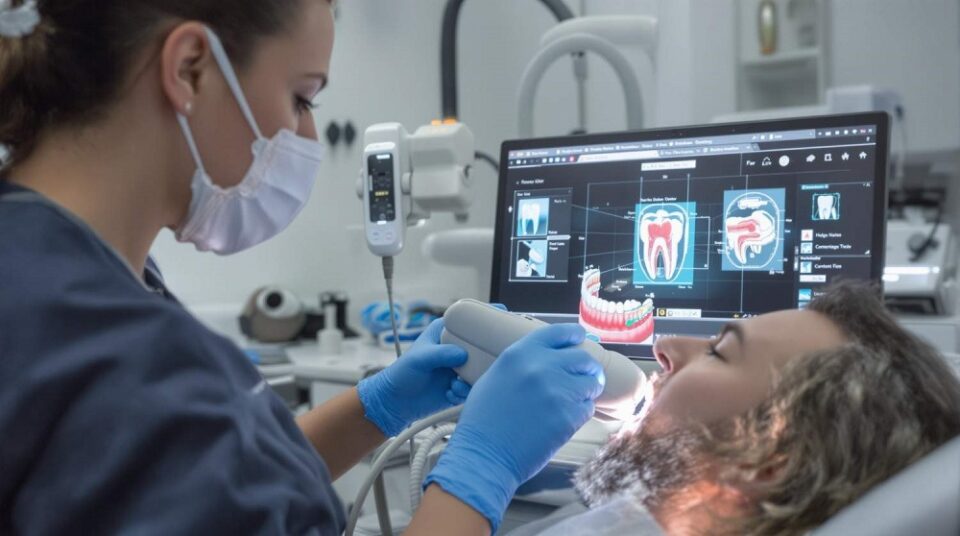
Modern clinics rely on intraoral scanners to capture dental anatomy. These handheld wands utilize advanced optical technology to capture thousands of three-dimensional data points within the oral cavity in seconds.
Digital impression systems offer distinct clinical advantages:
-
Elevated Patient Comfort: The small, ergonomic wand moves smoothly over the teeth, eliminating the mess, bad taste, and gagging associated with traditional trays.
-
Superior Accuracy: Digital scans eliminate variables that cause distortion in physical molds, such as material shrinkage, air bubbles, or tearing.
-
Instant Visualization: The real-time 3D model appears on an adjacent monitor immediately, allowing dentists to evaluate preparation areas instantly and show patients their exact oral structures.
-
Streamlined Communication: Digital files are sent instantly to dental laboratories via secure networks, reducing shipping times and accelerating overall production schedules.
Advanced Diagnostic Imaging
Accurate diagnostics form the foundation of successful dental treatment planning. Standard two-dimensional bitewing radiographs remain highly useful, but they offer a limited view because they flatten three-dimensional anatomical structures into a single plane. This can occasionally mask underlying pathology or distort root spatial relationships.
Cone Beam Computed Tomography has significantly upgraded diagnostic capabilities for complex dental cases. Unlike conventional medical CT scanners, which expose patients to higher radiation levels and require large machines, a dental cone beam scan uses a rotating gantry to emit a cone-shaped X-ray beam. The system captures hundreds of high-resolution images during a single low-dose rotation around the patient’s head.
The resulting dataset allows clinicians to view cross-sectional slices of the jawbone, trace nerve pathways, evaluate sinus cavities, and analyze the roots of the teeth with immense structural clarity. This technology is vital for planning dental implant placements safely, executing complex root canal therapies, and diagnosing impacted teeth or jaw joint disorders without guesswork.
Digital radiography has largely replaced traditional film X-rays across the industry. By using high-sensitivity electronic sensors instead of photographic film, modern dental offices have reduced patient radiation exposure by up to eighty percent. Furthermore, digital images require no chemical processing, appearing instantly on computer screens where dentists can magnify, contrast-enhance, and color-code the images to detect micro-cavities at their earliest stages.
Same-Day Restorative Systems
The traditional workflow for receiving a dental crown, veneer, or inlay required multiple appointments spread across several weeks. A patient would undergo tooth preparation, sit through a physical impression, wear a fragile temporary acrylic restoration for two weeks, and return for a second appointment to have the final restoration fitted and cemented.
Computer-Aided Design and Computer-Aided Manufacturing systems have compressed this multi-week sequence into a single clinical visit. Commonly referred to as same-day dentistry, this system integrates an intraoral scanner, specialized design software, and an in-office milling machine.
Once the dentist prepares the damaged tooth, they scan the area to create a digital model. The dentist then uses the software to custom-design the shape, margins, and contour of the new ceramic crown to match the surrounding teeth perfectly.
Once the design is finalized, the data is sent to an on-site milling unit or 3D printer. The machine carves the restoration from a solid block of high-strength lithium disilicate or zirconia ceramic in roughly fifteen minutes. The clinician stains, glazes, and fires the crown in a compact laboratory oven before bonding it permanently to the patient’s tooth. This eliminates the need for temporary crowns and secondary local anesthesia injections.
Minimally Invasive Laser Applications
The mechanical dental drill is a primary source of dental anxiety due to its high-pitched vibration and heat generation. Dental lasers have emerged as a highly effective alternative for a wide range of hard and soft tissue procedures.
Lasers emit a highly concentrated, specific wavelength of light energy that interacts precisely with biological tissues. In hard-tissue applications, lasers can target water molecules within tooth decay, vaporizing the damaged structure while leaving the healthy surrounding enamel and dentin completely untouched. Because lasers do not generate mechanical friction or heat, many minor cavity preparations can be completed without local anesthesia injections.
For soft-tissue procedures, lasers provide unmatched clinical precision. Periodontal specialists utilize dental lasers to remove diseased gum tissue, sterilize deep periodontal pockets, and reshape uneven gum lines. The laser energy cauterizes blood vessels and seals nerve endings simultaneously as it cuts. As a result, patients experience minimal bleeding during the procedure, reduced swelling, and significantly faster healing phases compared to traditional scalpel-based surgeries.
Artificial Intelligence in Clinical Practice
Artificial intelligence has evolved past theoretical research to become a highly functional diagnostic aid in modern dental practices. Sophisticated AI algorithms assist dentists by acting as an automated second pair of eyes when evaluating diagnostic images.
These software platforms are trained on massive datasets containing millions of clinical radiographs. When a dentist uploads a patient’s digital X-rays, the AI automatically analyzes the pixels to identify subtle density variations that signify early-stage bone loss, root fractures, or hidden decay beneath existing fillings.
By highlighting these areas on the screen, AI helps minimize diagnostic oversight and provides objective validation for recommended treatments. Patients gain greater confidence in their treatment plans when they see clear, objective data supporting their dentist’s observations.
The Broad Impact on Patient Experience
While these technological components are individually impressive, their collective value lies in how they reshape the overall patient experience. Dental visits have shifted toward an transparent, educational, and collaborative process.
When patients see high-definition 3D models of their teeth on a screen, understand their bone structure via clear digital imaging, and watch their custom restorations created in real time, their anxiety decreases. The reduction in physical discomfort, fewer required office visits, and highly predictable treatment outcomes encourage individuals to maintain regular dental care, ultimately preventing major oral health complications before they can develop.
Frequently Asked Questions
Are digital X-rays safe for pregnant patients?
Digital X-rays emit exceptionally low levels of radiation, making them highly safe. When a dental emergency or urgent diagnostic need arises during pregnancy, the use of digital radiographs combined with proper lead apron shielding over the abdomen and thyroid pose virtually zero risk to the developing fetus. Routine preventative X-rays are still typically deferred to the postpartum period as a standard precaution.
Why do some cavities still require a traditional drill instead of a laser?
While hard-tissue lasers are excellent for removing small to medium areas of decay, they cannot be used to remove old metal amalgam fillings or prepare teeth for large dental bridges. Drills are also required to shape and polish restorations rapidly. Dentists determine the tool selection based on the specific material constraints and structural requirements of each cavity.
How long do same-day ceramic crowns last compared to lab-made crowns?
Same-day crowns manufactured using in-office milling systems display identical structural longevity and durability to restorations made in a commercial laboratory. Both methods utilize high-grade, biocompatible ceramic materials like lithium disilicate and zirconia. With diligent oral hygiene and regular clinical cleanings, these restorations routinely last between ten and fifteen years.
Can intraoral scanners detect oral cancer?
Intraoral scanners are designed specifically to capture the three-dimensional shapes and contours of hard and soft tissues for restorative purposes rather than identifying cellular pathology. However, many modern intraoral cameras and specialized diagnostic wands use fluorescence technology to emit light wavelengths that highlight abnormal tissue changes beneath the surface, aiding early oral cancer screenings.
Does insurance cover treatments performed with advanced technologies?
Dental insurance coverage is based primarily on the specific procedure code rather than the technological tool used to complete it. For example, insurance covers a standard crown or a cavity preparation regardless of whether it was performed with a digital scanner, a traditional impression tray, a laser, or a mechanical drill. Patients do not typically incur specific surcharges for the use of advanced equipment.
How does AI protect the privacy of my dental records?
Artificial intelligence diagnostic platforms operate within strict regulatory frameworks, including federal health privacy laws. Patient data uploaded to AI networks is stripped of direct personal identifiers, such as social security numbers or home addresses, encrypting the medical images securely to ensure compliance with modern data safety protocols.
